When it comes to a healthy heart, getting the right test at the right time can make all the difference. But with so many options available, the big question is: Echo test vs. ECG — which one do you actually need? While both tests play a crucial role in diagnosing heart conditions, they serve very different purposes.
Understanding these differences can help you take the right steps toward better heart health. Here are some stats to know:
- According to the World Health Organization, cardiovascular diseases (CVDs) are responsible for an estimated 17.9 million deaths annually.
- What’s even more concerning is that, as per Harvard Health, approximately 45% of all heart attacks are “silent,” meaning they occur without noticeable symptoms.
- Additionally, the World Heart Federation reports that deaths from cardiovascular diseases have surged by 60% over the past 30 years, rising from 12.1 million in 1990 to 20.5 million in 2021.
With heart-related conditions becoming increasingly common, knowing the difference between an Echo test and an ECG test can help you make informed decisions about your health.
Let’s break down how each test works; when you might need them; and how they compare – so you never have to second-guess your next heart check-up.

What is an Echocardiogram and How Does it Work
An echocardiogram, or Echo test, is a painless imaging procedure that helps evaluate heart structure and function. Essentially, it uses high-frequency sound waves to create live images of the heart. As a result, this test helps doctors check heart health and detect abnormalities early. It helps in the following way:
- Diagnosing heart disease – Helps detect heart conditions before symptoms worsen.
- Checking heart function – Assesses how well the heart pumps blood.
- Identifying abnormalities – Detects valve diseases, blood clots, and weak heart muscles.
How it Works:
The Echo test vs. ECG debate arises because both tests check heart health, yet they work differently. An echocardiogram uses ultrasound waves to capture detailed heart images. Specifically, the device sends sound waves, which bounce off the heart and return as echoes. Then, a computer processes these echoes to create real-time visuals.
- Non-invasive procedure – No needles or incisions are required, making it a comfortable option.
- Real-time imaging – Helps assess heart chambers, valves, and blood flow with high accuracy.
- Quick and safe – Usually takes 30-60 minutes, and importantly, there’s no radiation exposure.
Since an Echo test provides live images, it helps doctors detect structural issues efficiently. In fact, this is a major differentiator in the Echo test vs. ECG comparison.
Echo test vs. ECG: Different Types of Echo
The Echo test vs. ECG debate continues because each test serves a unique purpose. An echocardiogram has different types, each designed for a specific heart condition. For this reason, understanding these variations helps determine which test you might need for an accurate diagnosis.
- Transthoracic Echocardiogram (TTE)
A Transthoracic Echocardiogram (TTE) is the most commonly used echocardiogram. It involves placing a probe on the chest to capture the heart’s images. Since it is completely non-invasive, there is no discomfort or special preparation needed.
Moreover, it provides detailed visuals, helping doctors assess heart chambers, valves, and overall function effectively. This makes it a widely-discussed subject during the Echo test vs. ECG debate.
- Transesophageal Echocardiogram (TEE)
A Transesophageal Echocardiogram (TEE) is used when clearer images are needed, especially for detecting blood clots and valve issues. In this procedure, a small probe is passed down the throat, providing a closer look at the heart.
Since mild sedation may be required, it is safely performed under medical supervision. This gives it an edge in comparison to the Echo test or ECG.
- Stress Echocardiogram
A Stress Echocardiogram is conducted before and after exercise to evaluate the response of the heart to stress. It is particularly useful for detecting coronary artery disease by identifying reduced blood flow to the heart muscles.
As an alternative to treadmill stress tests, it benefits individuals unable to perform physical activity. When considering the Echo test or ECG, a Stress Echo offers a more dynamic assessment of the heart function.
- Doppler Echocardiogram
A Doppler Echocardiogram measures blood flow in the heart, helping identify abnormalities in circulation. It is essential for diagnosing valve disorders by detecting leaking or narrowed valves.
Moreover, it can be combined with other echocardiogram types to provide additional insights into heart function and overall cardiovascular health. This further highlights how the Echo test vs. ECG serves different diagnostic purposes.
- 3D Echocardiogram
A 3D Echocardiogram creates a detailed three-dimensional image of the heart, allowing doctors to plan surgeries and treatments more effectively. It is more precise than traditional echocardiograms, making it useful for diagnosing complex heart conditions.
Additionally, it is often combined with TTE or TEE to provide better insights into structural defects.
Each type of Echo has its own purpose; however, they all help evaluate heart health.
What is an Electrocardiogram and How Does It Work?
As we move forward with the debate of the Echo test vs. ECG, let’s learn about ECG. An electrocardiogram (ECG) is a simple, non-invasive test that records the heart’s electrical activity. It helps detect irregular heartbeats, blockages, and overall heart health. It helps in:
- Identifying heart rhythm problems like arrhythmia;
- Detecting previous or ongoing heart attacks;
- Diagnosing coronary artery disease; and
- Monitoring heart health after surgery or treatment.
How it Works
Now, let’s break down how an ECG works:
- Electrodes are placed on the chest, arms, and legs;
- These electrodes detect electrical signals from the heart; and
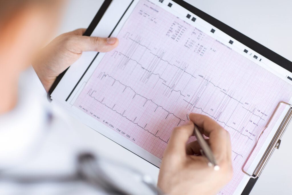
- A machine records the heart’s activity as a graph.
Since ECG results focus on electrical activity, they provide insights that an echocardiogram cannot.
Echo test vs. ECG: Different Types of ECG
ECG tests come in various forms, each serving a specific purpose. Let’s explore the different types of ECG and their uses. This fundamental difference defines the Echo test vs. ECG comparison.
- Resting ECG:
A Resting ECG is the most common type performed while the patient is lying still. Electrodes are placed on the chest, arms, and legs to record the heart’s electrical activity. Since no movement is involved, it provides a clear baseline reading.
Doctors use it to detect arrhythmias, heart attacks, and other abnormalities at rest.
- Stress ECG:
A Stress ECG monitors heart activity during physical exertion. The patient walks on a treadmill or pedals a stationary bike while electrodes record heart signals. This test helps detect coronary artery disease and assess blood flow under stress.
It is particularly useful for identifying conditions that may not appear during a resting ECG.
- Holter Monitor ECG:
A Holter Monitor ECG records heart activity over a period of 24 hours to 48 hours. A small portable device continuously tracks the heart’s electrical signals. This test helps detect irregular heart rhythms that occur sporadically.
Patients go about their daily activities while wearing the monitor, providing a more accurate assessment of the heart function.
- Event Monitor ECG:
An Event Monitor ECG is used for patients experiencing occasional symptoms like dizziness or palpitations. Unlike a Holter monitor, this device is worn for weeks or even months. It records heart activity only when activated by the patient or automatically during irregular heartbeats.
This method helps diagnose intermittent arrhythmias that may not appear in short-term tests.
- Signal-Averaged ECG:
A Signal-Averaged ECG provides a detailed analysis of the heart rhythms by averaging multiple ECG recordings. It helps detect subtle electrical abnormalities that may indicate a risk of serious arrhythmias. This test is often used for patients with unexplained fainting episodes or those at risk of life-threatening heart conditions.
Now that we’ve explored these variations, let’s compare the Echo test vs. ECG to understand which one is right for specific heart concerns.

How are ECG and Echo Tests Done?
The Echo test vs. ECG debate often comes up when doctors need to assess the heart function. While both tests help evaluate heart health, they work in entirely different ways. Understanding the procedures can help you feel more prepared. Now, let’s explore how each test is done.
- How is an ECG Done?
During the ECG procedure, small electrodes are carefully placed on the chest, arms, and legs. These electrodes detect electrical signals which are then displayed as a graph on a monitor for analysis.
Moreover, the test usually takes just 5 minutes to 10 minutes. Patients must remain still to prevent any interference in the readings. Since an ECG involves no radiation or discomfort, it is considered a safe and routine test for diagnosing heart rhythm issues.
- How is an Echo Test Done?
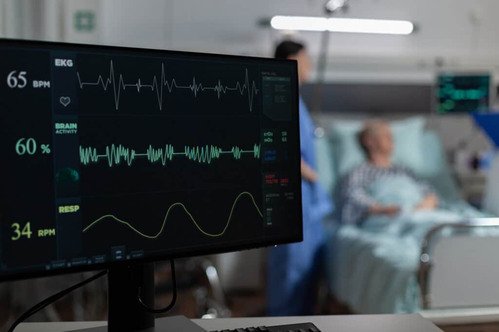
Unlike an ECG, an Echo test creates moving images of the heart using high-frequency sound waves. A technician first applies gel to the chest and then moves a handheld device over the skin. This sends sound waves that bounce off the heart, generating real-time visuals on a monitor.
Additionally, the procedure typically lasts between 30 minutes and 60 minutes. Patients may need to shift positions to allow for better imaging. Unlike an ECG, an Echo provides highly detailed visuals of heart structures, making it more effective for detecting valve diseases and muscle function issues.
- Key Differences in the Testing Process
On the one hand, an ECG focuses solely on electrical activity. On the other, an Echo captures live heart images. The former is much quicker while the latter provides more in-depth details.
Since both tests serve different purposes, doctors often use them together for a complete heart assessment, further enriching the Echo test vs. ECG comparison.
When Do You Need ECGs and Echo?
The comparison of Echo test and ECG is crucial because each test plays a different role in assessing the health of the heart. While both are essential, they serve distinct purposes. Understanding when to get each test allows you to make informed decisions for early diagnosis and better treatment.
- When Do You Need an ECG?
An ECG is recommended when doctors suspect heart rhythm issues. It records heartbeat patterns and detects irregularities. If you have palpitations, an ECG helps check for arrhythmia. Chest pain or discomfort may indicate a heart attack or blocked arteries.
An ECG helps identify these conditions. Shortness of breath or dizziness could signal heart rhythm disorders, making an ECG necessary.
ECGs are also part of routine check-ups. People with a family history of heart disease often need this test. Since it is quick and painless, doctors use it widely for initial heart screenings.
- When Do You Need an Echo?
Unlike an ECG, an Echo test provides detailed images of the heart. It helps diagnose structural problems and assess the heart function. Doctors recommend it when they suspect issues with heart muscles or valves.
If a doctor hears a murmur, an Echo checks for valve narrowing or leakage. Symptoms like swelling, fatigue, or breathlessness may indicate heart failure, requiring an Echo for confirmation. After a heart attack, this test helps assess muscle damage and blood flow.
An Echo is also useful for monitoring existing conditions. Patients recovering from heart disease or undergoing treatment often need regular Echo tests to track progress and detect changes early.
What are the Differences Between an Echo test and ECG?
The debate often arises when doctors need to assess heart health. While both tests evaluate the heart, they serve different purposes. Therefore, understanding the differences between the Echo test and ECG helps determine which test suits your condition best.
- Purpose and Function:
An ECG focuses on the heart’s electrical activity. It detects irregular heartbeats, heart attacks, and conduction problems. In contrast, an Echo test provides moving images of the heart. As a result, it helps diagnose structural issues, valve disorders, and heart muscle function.
- Procedure and Time Taken:
An ECG is a quick test that takes around 5 minutes to 10 minutes. Small electrodes are placed on the skin to record heart rhythms. Meanwhile, an Echo test takes 30 minutes to 60 minutes. A handheld device uses sound waves to capture real-time heart images.
- Key Uses in Diagnosis:
Doctors use ECGs to check for arrhythmia, heart attack signs, or unexplained chest pain. Additionally, it helps monitor heart conditions over time. On the other hand, an Echo helps detect heart failure, valve disease, and congenital defects.
Consequently, each test plays a vital role in diagnosing different heart conditions.
- Safety and Risk Factors:
Both tests are safe and painless. An ECG does not use radiation, making it a risk-free procedure. Likewise, an Echo relies on sound waves to create heart images. However, transesophageal echocardiograms (TEE) may require mild sedation for better image clarity.
Since both tests provide unique insights, doctors may recommend both ECG and Echo for a complete heart assessment. Now, let’s explore when you might need each test.
Side Effects of ECGs and Echo
When comparing the Echo test and ECG, safety is a key factor. Both tests are generally risk-free, but some minor side effects might appear. Learning about these can help you approach the test with confidence.
- Possible Side Effects of ECG:
An ECG is a non-invasive test as it does not involve needles or radiation. However, some minor effects may still occur. The adhesive from electrodes may cause mild skin irritation leading to slight redness or itching.
Some patients also feel some discomfort when removing the electrodes, especially if they have sensitive skin.
In some cases, nervousness during the test may result in temporary dizziness or anxiety. This reaction is more common in patients who feel stressed about medical procedures. Fortunately, these effects disappear quickly and do not require any treatment.
Since an ECG only records electrical signals, it does not affect the heart or body directly.
- Possible Side Effects of Echo:
An Echo test is also safe, but certain types may have minor side effects. Many patients experience a cool sensation on the skin due to the gel applied before the test. The pressure from the handheld probe may also cause slight discomfort in the chest, especially if deeper imaging is required.
For those undergoing a stress Echo, temporary dizziness may occur after exercise. This reaction happens due to increased heart rate and physical exertion. In a transesophageal echocardiogram (TEE), mild sedation is often necessary.
- Are These Side Effects a Concern?
Both ECG and Echo are low-risk tests with minimal side effects. Most discomfort is temporary and does not require medical attention. Since these tests help detect heart conditions early, their benefits far outweigh minor effects.
Understanding these possibilities can help patients feel more at ease before undergoing either test.
Conclusion: Echo test vs ECG
To conclude, the comparison between the Echo test and ECG is essential for understanding heart health evaluations. While an ECG measures electrical activity, an Echo creates moving images of the heart. Since both tests serve different purposes, they help doctors diagnose a wide range of heart conditions effectively.
If you experience chest pain, irregular heartbeats, or shortness of breath, your doctor may recommend an ECG, an Echo, or both. Understanding the differences between the Echo test and ECG can help you make informed choices about your heart health.
Your heart health should never be ignored. At Riverside B&J hospital, we provide expert ECG and Echo testing with precision and care. Our experienced team ensures a stress-free experience with accurate results. Book your appointment today and take control of your heart health!
FAQs about Echo test vs ECG
- Which is More Accurate, ECG or Echo?
Both tests serve different purposes. An Echo test vs ECG comparison depends on whether electrical activity or heart structure needs evaluation.
- Is ECG Required When Echo is Normal?
Even if an Echo test is normal, an ECG may still be needed to check heart rhythm abnormalities.
- What are the Symptoms of a Blocked Artery?
Chest pain, shortness of breath, and fatigue may indicate blocked arteries, requiring an Echo test vs ECG for diagnosis.
- Are Echo Tests and ECG Tests Safe?
Yes, both Echo test vs ECG tests are safe, painless, and non-invasive with minimal side effects.
- Can an Echo Test or an ECG Test Be Done at Home?
Portable ECG devices are available, but a clinical Echo and ECG test provide more accurate and detailed results.

