
Introduction
CT Angiography in Navi Mumbai
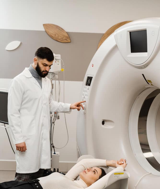
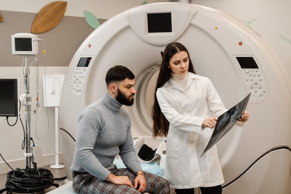
An angiography in Navi Mumbai is a non-invasive cardiac CT scan where Dr. Vinay Jaiswal visualizes coronary arteries using contrast dye and detailed X-ray images for 3D assessment.
Meet Our Specialist
Dr. Vinay Jaiswal - M.D. D.M. Cardiology
Meet Our Specialist
Dr. Pritish Bagul - M.D. D.M. Cardiology
Meet Our Specialist
Dr. Jui Bagul - M.D. D.M. Neurology
About
What is angiography?
Angiography is an X-ray exam used to diagnose blockages and abnormalities in arteries, veins, and blood flow.
Performed by an interventional radiologist, it identifies narrowed arteries, heart muscle issues, or valve abnormalities in the heart muscle.
When is it used?
Coronary Artery Disease (CAD)
Angiography detects blockages or narrowings causing chest pain or heart attacks.
Peripheral Arterial Disease
Evaluates blood flow and detects blockages causing extremity pain or ulcers.
Pulmonary Arterial Disease
Pulmonary angiography assesses lung vessels for embolism, hypertension, or abnormalities.
Cerebral Vascular Disease
Cerebral angiography identifies brain vessel abnormalities, aneurysms, or arteriovenous malformations.
Renal Artery Disease
Renal angiography examines kidney vessels, identifying blockages causing hypertension or affecting function.
What happens during the procedure?
Catheter Insertion
A catheter is inserted into an artery through a small incision.
Contrast Dye Injection
Contrast dye highlights blood vessels on X-ray to detect abnormalities.
Continuous Monitoring
Heart, blood pressure, and oxygen levels monitored during the procedure.
Blockage Treatment
Angioplasty or thrombolysis performed if blockages or narrowing are identified.
Non-Invasive Alternatives
CT angiography provides detailed images without catheter insertion for certain cases.
Cost of Angiography in Mumbai
On average, angiography in Mumbai costs between ₹8,000 - ₹2,00,000.However, the total cost may vary depending upon the type of procedure performed, facility type, physician’s expertise, procedural inclusions, and insurance coverage, if any.
Common angiography types include:
Common angiography types include:
Angiography Type | Estimated Cost |
Conventional Coronary | ~ ₹35,000 |
Coronary Angiography | ~ ₹8,000 – ₹2,00,000 |
CT Angiography | ~ ₹14,000 |
Peripheral Angiography | ~ ₹24,500 |
Pulmonary Angiography | ~ ₹8,500 |
Renal Angiography | ~ ₹10,500 |
Digital Subtraction Angio | ~ ₹8,500 |
MRI Angiography | ~ ₹20,500 |
Cerebral Angiography | ~ ₹25,000 – ₹31,000 |